Surgery Revision Day MBBS 2015
Yesterday we had Surgery Revision Day for MBBS. It started at 8.30am and ended at 1.00pm. We gathered at Lecture Hall 3 JHC first, and then went to separate rooms in Exam Room.
We were divided into 9 groups and there were 9 stations altogether, 20 minutes allocated for each station. I was in Group 6, with my other 13 friends.
Here's a summary on what each station is all about.
STATION 6
Lump and Bump - Dr. Yusuf
Lipoma Lipoma Lipoma. Please come out in exam lol
A middle aged man, comfortably sitting on a chair. On inspection, there is an obvious swelling just below left scapula. There is no overlying skin changes, not erythematous, no punctum, sinus opening or discharge.
On palpation, the size is 13 x 10 cm, oval in shape, has well-defined margin, soft in consistency, smooth surface, diffuse swelling, mobile, non-tender, not warm, not attached to skin or muscles, dull on percussion, transillumination test negative.
On palpation, the size is 13 x 10 cm, oval in shape, has well-defined margin, soft in consistency, smooth surface, diffuse swelling, mobile, non-tender, not warm, not attached to skin or muscles, dull on percussion, transillumination test negative.
Provisional diagnosis : Back lipoma (fat accumulation in subcutaneous tissue)
Ddx:
1. Sebaceous cyst (collection of sebum in a sac located in dermis or subcutaneous tissue)
Points against : sebaceous cyst has punctum, non mobile and attached to skin
2. Abscess (collection of pus in a confined space)
Points against : abscess is erythematous, warm and tender
3. Carbuncle (subcutaneous tissue necrosis with multiple sinus opening)
Points against : carbuncle is hard and tender, with sinus opening
STATION 7
Surgical Instruments - Mr. Chan
This station was a bit difficulttt. Must know the name of instrument, how to use it, indications and contraindications.
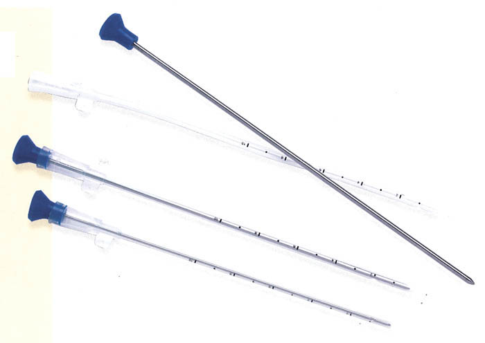
1. Trucut biopsy needle
 |
| The needle Image taken from this website |
 |
| Principle of trucut biopsy Image taken from this website |
2. Sigmoidoscope
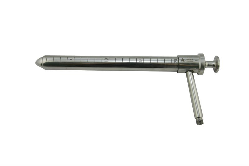 |
| The scope Image taken from this website |
 |
| Different types of endoscopes Image taken from this website |
3. Triple lumen Foley catheter
The three-way tubing has an outlet for drainage of urine, an inlet for water inflation of the balloon, and an inlet for normal saline used for irrigating the urinary system most commonly for gross hematuria.
 |
| Different types of foley catheters Image taken from this website |
4. Chest tube
 |
| The tube and trocar inside Image taken from this website |
 |
| How to place the chest tube Image taken from this website |
| Safety triangle Image taken from this website |
5. Skin stapler
Advantage is faster, less scar. Disadvantage is that it's expensive!
 |
| The stapler Image taken from this website |
6. Last question, Mr. Chan asked us - what is this?
 |
| Image taken from this website |
It's actually the cover of chest tube lol that was funny
STATION 8
X-Ray - Dr. Hisham
We did not have time to go through all xrays, basically we revised on how to read xrays.
Case 1
65 year-old gentleman with history of myocardial infarction 5 years ago, did angioplasty, on beta blocker and frusemide, presented with 5 days history of worsening dyspnea. Interpret his chest xray.
This is the chest xray of Mr.S, taken on (date) at (time), AP view (portable), right side is marked correctly. The patient is rotated, however it has good penetration and exposure. There is homogenous opacity over the right lung with blunting of costophrenic angle.
Diagnosis - Right pleural effusion
How to assess rotation, penetration and exposure?
1. Rotation : look at the distance between clavicle and spinous process (teardrop shape). It must be the same between right and left side.
2. Penetration : look at the last few lumbar vertebrae, near the heart, they must be visible
3. Exposure : can see first rib, diaphragm and lateral border of ribs. Anterior axillary line must intersect the middle of 6 1/2 anterior ribs.
How to manage this patient?
ABCDE first! Remember to do step by step, insert 2 large bore branula, take blood for investigations, CXR, ECG, and then chest tube insertion.
Case 2
65-year-old man, psychiatric patient, bed bound, presented with recurrence abdominal pain, abdominal distension and constipation. Interpret his abdominal xray.
Coffee bean sign!
Diagnosis - Sigmoid volvulus
Management?
ABCDE! Branula, hydration, consent for immediate sigmoidoscopy to derotate sigmoid. Then, put rectal tube. After that, can decide for rectopexy or sigmoid colectomy.
Complications of sigmoid volvulus?
Can cause bowel obstruction, ischaemia, perforation.
Case 3
Barium swallow : bird-beak appearance
Diagnosis? Achalasia!
Case 4
KUB XRay : Right staghorn calculi!
STATION 9
CT Scan - Prof. Saufi
We were given 5 minutes to discuss on the findings, and then Prof came in to discuss.
Case 1
CT scan of thorax and abdomen, non contrast, axial view, soft tissue window. There are multiple hypodense lesions in liver, irregular margin. Spleen and kidneys are normal. Right pleural effusion noted in right lung.
Ddx for hypodense lesion in liver?
Liver abscess, liver metastasis or liver carcinoma
Your provisional diagnosis?
Liver carcinoma with lung metastasis
What investigation you want to do next?
Contrast CT scan of thorax and abdomen
Case 2
CT scan of brain, non contrast, axial view, soft tissue window. There is a heterogenous lesion lesion of mixed density, crescent shape, noted at right temporo-parietal region. Shifting of midline structures noted with left ventricle dilatation.
Impression?
Acute on chronic right subdural haematoma
What are the symptoms and signs patient can come with?
Altered consciousness, vomiting, seizure, dilated right pupil, left hemiparesis
STATION 10
Pathology Pictures - Dr. Azim :)
Dr. Akmal Azim is our new lecturer - and he is a plastic surgeon! So cool!
1. Specimen of thyroid gland with multiple nodules on right and left lobe.
Diagnosis?
Multinodular thyroid
Investigations?
Blood (TFT, FBC), Imaging (US neck) and Special tests (FNAC)
Management?
Medical (carbimazole), Surgical (thyroidectomy) and Radioiodine therapy
2. Endoscopic view of pedunculated and sessile polyp
Symptoms patient can have?
Alteration in bowel habit, per rectal bleeding, symptoms of obstruction
Any special condition/disease you know that is related to this picture?
- Hereditary Non Polyposis Colorectal Cancer (HNPCC)
- Familial Adenomatous Polyposis (FAP)
What to do if patient has one of these condition?
Patient needs regular surveillance as there is risk of malignant change, need to screen family members
3. Specimen of large intestine showing multiple outpouchings with gangrenous area
Diagnosis?
Diverticular disease or diverticulosis
Cause?
Reduced fiber intake, predispose to constipation and straining, recurrence leads to weakening of colon wall and then outpuching
Symptoms patient can have?
Usually patient comes with pain, rarely per rectal bleeding.
Complications?
If the area becomes inflamed and infected, can cause abscess formation. Can also cause perforation and fistula.
4. Middle aged man with scleral jaundice
Differential diagnosis?
- Pre hepatic : haemolytic anemia, G6PD
- Hepatic : hepatitis, alcoholic liver disease, chronic liver disease
- Post hepatic : bile duct stone, stricture, head of pancreas tumour, Mirizzi syndrome
Risk factors of gallbladder stone?
Female, Forty, Fat, Fertile
5. Specimen of gallbladder and pigmented gallstones
Types of gallstones?
Cholesterol (most common), pigmented, mixed
STATION 1
Abdomen - Mr. Mizam
Who wants to volunteer to examine this patient's abdomen?
Middle aged Chinese man comfortably lying in supine position. He looks pale but not jaundiced. On inspection of abdomen, there is no surgical scar seen, no dilated veins, visible pulsation or stigmata of chronic liver disease (spider naevi, gynaecomastia, loss of axillary hair, caput medusae). There is no inguinal hernia.
On palpation, there is a mass located at left iliac fossa measuring (? cm) that has well-defined margin, round in shape, can get above and can get below, smooth surface, firm in consistency, not mobile, tender on palpation, dullness on percussion (don't percuss if patient is in pain), not attached to skin or muscles. There is no hepatosplenomegaly, kidneys were not ballotable, no ascites.
Points to remember:
1. Stand at the end of patient's bed for general inspection
2. Look at patient's face when palpating abdomen or mass
3. Ask patient to flex his neck to check if mass is below or above muscle
4. Do skin pinch to check for attachment to skin
We didnt manage to discuss about the diagnosis, investigation and management because the time's up. Our friend says that it's a case of rectosigmoid carcinoma.
STATION 2
Thyroid - Prof. Junaini
Must master thyroid examination, confirm will come out in exam! At that time, there was no real thyroid patient, so one of us had to examine a normal male with no thyroid enlargement.
1. Ask patient to swallow water and protrude tongue
2. Do thyroid examination from back of patient
3. Check trachea and percuss retrosternal, check lymph nodes
4. Auscultate carotid bruit, check Pemberton sign
5. Check peripheral - fine tremor, sweating palms, pulse for atrial fibrillation, eyes for ophthalmopathy, shoulder for proximal myopathy
What do you know about thyroid carcinoma?
 |
| I made this myself, for future reference. Some of the information is from the internet. |
STATION 3
Breast - Mr. Faiz
Practice, practice, practice! No patient with breast lump, so one of us had to wear fake boobs to be examined lol
Organise your examination:
1. Inspection - look for asymmetry, skin dimpling, peau de orange, discharge etc.
2. Ask patient to raise both hands (look if there is anything underneath breast). Ask patient to press hands on hips (if there is attachment to pectoralis major, mass will become prominent).
3. Examine normal breast first, then the pathological one. Cover the other breast.
4. Ask patient to raise hand above head.
5. Lift up breast and look under breast for surgical scar (breast augmentation)
6. Palpate four quadrants + on nipple + below scapula
7. If there is a mass, describe it as usual - site, size, shape, surface etc.
8. Don't squeeze the nipple! Ask patient if she has any nipple discharge, if not then proceed. If yes, ask if she can demonstrate.
9. After examine both breasts, ask patient to sit down facing you. Check for axillary lymph nodes. Anterior, apical, medial, lateral and posterior. Check cervical and supraclavicular lymph nodes. Check spine tenderness (bone mets), percuss lung (pleural effusion, lung mets), auscultate lungs (reduced air entry, crepitations)
10. Ask patient to lie supine. Check for hepatomegaly.
STATION 4
Hernia - Mr. Islah
The patient was very willing to let us examine him, one person per group, that's a total of 9 students touching him....thank youu pakcik may Allah bless you!
Elderly man, lying comfortable in supine position. On inspection of abdomen, there is a midline laparotomy scar noted, with two transverse scars at right and left iliac fossa. There is an obvious right inguinal swelling, not extending to scrotum. There is no erythematous changes, no skin excoriation, no dilated veins, no punctum or discharge, no visible pulsation or peristalsis. Cough impulse is positive as the mass becomes obvious after coughing.
On palpation, mass is oval in shape 5 x 4 cm, soft and doughy, has well-defined margin, non tender, not warm to touch, can get below and reducible. Occlusion test is negative.
Genitalia examination is normal. Scrotum is well-developed. Both testis palpable, normal size. Normal spermatic cord, no bag of worms felt.
Since patient's swelling is completely reducible, I would like to ask patient to stand for further assessment. When patient standing, do transillumination test. It's negative.
I would like to complete my examination by doing per rectal examination, abdomen and respiratory examination.
How to do occlusion test?
Make sure swelling is completely reducible. Find landmark. Midway between ASIS and pubic tubercle (first bony prominence after pubic symphysis). Occlude opening with one finger. Ask patient to cough. If negative, ask patient to stand, finger still occlude the opening. Ask patient to cough again.
Your complete provisional diagnosis?
Right recurrent completely reducible direct inguinal hernia with no complications such as strangulation, ischemia or incarceration.
How to know if its bowel or omentum?
- Bowel : Visible peristalsis on inspection, gurgling sensation on palpation and when you try to reduce it, initially it's hard and then becomes easy, bowel sound on auscultation
- Omentum : No visible peristalsis, soft and doughy on palpation and when you try to reduce it, it's easy initially but hard at last, no bowel sound heard
How to manage this patient?
Since this is a recurrent disease, I would like to take complete history and assess his risk factors (occupation, congenital, complications of previous operation, abdominal mass, urinary and bowel symptoms, respiratory problems, heavy lifting). After that, I would like to perform complete physical examination. Then, do pre-op assessment, optimize patient's condition, take blood for investigations, do CXR, ECG. Prepare patient for laparoscopic hernioplasty where we put in mesh to induce fibrosis and prevent hernia.
Advantages of laparoscopic surgery?
Small incision, less bleeding, less infection, faster recovery, reduced hospitalisation, reduced chronic pain
Disadvantages?
Need highly-experienced surgeon, longer operating time, risk of recurrence if surgeon is not experienced enough
Other options for surgery?
- Herniotomy : excision of sac after reduction, usually in children because they have weak and immature ligaments.
- Herniorraphy : reconstruction by using patient's own tissues
Type of open repair surgery that you know of?
- Lichtenstein : most common, flat mesh is placed on top of the defect
- Shouldice : four-layer reconstruction of fascia transversalis, difficult to perform
- Bassini : tension repair, edges of defect are sewn back together without any mesh
- Desarda : simpler, faster, tension-free, mesh-free
Read more about Inguinal Hernia Surgery in this website.
Special types of hernia that you know of?
- Sliding hernia : an organ is part of hernia sac eg. colon or urinary bladder
- Pantaloon hernia (saddle bag hernia) : combined direct and indirect hernia
- Maydl's hernia : two adjacent loops of small intestine are within a hernial sac with a tight neck - double lumen.
- Richter's hernia : hernia involving only one sidewall of bowel
- Littre's hernia : hernia involving Meckel's diverticulum
Read more about other types of hernia (there are a lot!) in this website.
STATION 5
Vascular - Mr. Ahmad Faidzal
The patient is a middle-aged Indian lady with varicose veins. We managed to feel defect in her veins. We did Doppler as well.
I have written about peripheral vascular examination last year, you can read it here. This is just a summary.
Remember : Inspection, Inspection, Inspection - Palpation, Percussion, Auscultation and Special Test.
Arterial system
1. Inspect for muscle wasting, colour, chronic signs of infection, arterial ulcer
2. Palpate for temperature, CRT, bone or joint tenderness, pulses (proximal to distal)
3. Ascultate bruit for AV malformation
4. Buerger's test - lift patient's leg and look at what angle it turns pale
5. Complete examination by doing ABSI, doppler, carotid pulse, cardiovascular, neurological and abdomen examination
What are the 6Ps of acute limb ischemia?
Pain, Pallor, Perishingly cold, Paralysis, Paraesthesia, Pulseless
Venous system
1. Inspect for calf swelling, lipodermatosclerosis, previous scar or healing ulcer, varicosity
2. Palpate the varicosity, tenderness (phlebitis), hardness (thrombosis), cough impulse
3. Auscultate bruit for AV malformation
4. Percussion is optional
5. Tradelenburg test and Multiple Tourniquet test
6. Complete examination by doing abdomen, per rectal, arterial system examination
Sooo... 10 stations COMPLETED!
Hope we can remember all of these points during exam!!
Hope all of us will pass the exam!! Amiiinnnn
source http://dizzyzizzy.blogspot.my/2015_06_01_archive.html?m=1